|
PART 1. Sep 24, 2011 The owner wanted a diagnosis from a male vet at Toa Payoh Vets. She had been to Vet 1 who referred her to Vet 2, but she said she still had to pay Vet 1 for a "non-diagnosis". I showed her that Vet 1 did diagnose "lipid" deposits in the right eye and then referred her to Vet 2. "It must have been a long time since I saw you," I said. I usually keep older records stored outside the Surgery. She showed me my 2006 Toa Payoh Vets receipt with my pen illustration of "left eye white central scarred ulcer - 3mm diameter". I was impressed that she kept records of her dog. It was some 5 years ago! "It was the left eye affected," she said. "The spot disappeared after a while." "Did you use the eye ointment?" I asked. "No," she said. "My dog disliked it. "Her ulcer disappeared without me using the eye ointment." It was good to meet up again and such reunions are happy incidents in veterinary practice as some children of my clients would have grown up to young adults or have married. EXAMINATION 1. To check whether the right eye had corneal ulcers or not in addition to the lipid deposits. I showed my assistant Min how I used the fluorets paper strip. I placed it onto the lower fornix and closed the eyelid. The dog blinked many times. Green dye gathered below the lower conjunctiva. "Is it dangerous to my dog's eye?" the owner asked. "No," I showed her the seller was Bausch and Lomb which she had instant brand recognition. She said: "There was a lawsuit against this company for the contaminated contact lens solution." But she knew I was using a reliable approved eye dye to use on her dog's eyes. Results: No green ulcers in the right eye but there were 11 to 1 o'clock ulcers in the left eye! 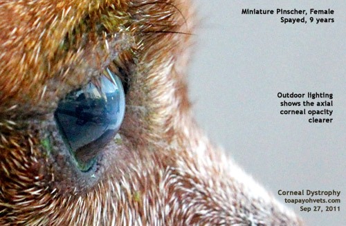 2. Digital photography useful esp. side views of
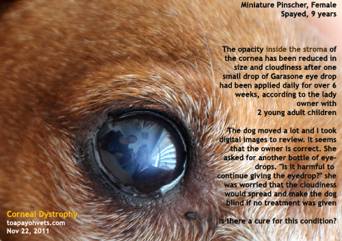
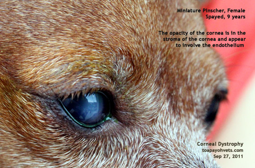
corneal degeneration. Axial central corneal
degeneration 4x3 mmx3mm. It looks like it is
going to bulge out and ulcerate. The Owner had said
the swelling suddenly appeared in the last
month. But I showed her that Vet 1 had recorded
that white deposits in the right eye had been seen in
the Sep 2010 visit. Only that the deposits were few. 2. Digital photography useful esp. side views of
corneal degeneration. Axial central corneal
degeneration 4x3 mmx3mm. It looks like it is
going to bulge out and ulcerate. The Owner had said
the swelling suddenly appeared in the last
month. But I showed her that Vet 1 had recorded
that white deposits in the right eye had been seen in
the Sep 2010 visit. Only that the deposits were few. 3. Blood tests for thyroid and cholesterol not done yet due to financial considerations. Hypothyroid and high cholesterol are said to be important in this degeneration where aqueous fluid enters the endothelium, causing edema and whiteness and swelling (side view of image). DIAGNOSIS Right eye - corneal endothelial dystrophy. No definite cure as this is likely due to aging. Blood tests for thyroid hormones and cholesterol levels advised. "Some old people get cholesterol lumps above their eyelids," I gave an analogy but she did not know what I meant as she had not seen them. Left eye - corneal ulcers due to eye rubbing of the 3 upper eyelid "melanomas". Excision of the melanomas will prevent ulcerations. TREATMENT Usually not curative. Low fat diet 8% fat? Check thyroid hormones and cholesterol levels. Prevent further corneal degeneration? E-collar and antibiotics to prevent left eye rubbing although the owner said she never saw actual rubbing. |
PART 2.
Monday, Sep 26,
2011
I review 47 images of the eyes of the fidgeting
Miniature Pinscher whose owner reject injections and
anaesthetics as a matter of personal preferences.
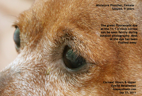
The green dye had stained the left eye 11-1 o'clock
cornea a zigzagging green. I saw it. The owner saw it.
But my camera techniques are lacking in that I can't
capture the ulcer clearly in the operating room after
application of the dye and doing hand-held. I had one
respectable image. What I needed was a reflector but
then Sunday was a busy day and I have not organised
myself.
This morning at 8.30 am, I went through the 47 images
again at home. Miniature Pinschers don't suffer from eye
ulcers unlike the Shih Tzus and the Pekineses. She did
have a small central eye ulcer scar of 3mm in diameter
in 2006 when the owner first saw me and that was the
only time I was consulted.
Clients in Singapore doctor hop whether it is human or
veterinary medicine but the vet must do his best in
the diagnosis and bedside manners as well as in his
receptionist services and other aspects. There are
many factors involved in retaining a client's loyalty.
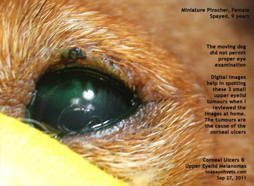
In any case, I was surprised that I had not seen the 3
melanomas on the upper eyelid at the 11-1 o'clock
position. That is the cause of the ulcer which
appeared just below these "melanomas." The 9-year-old
spayed female dog felt irritated by these 3x3 mm lumps
and must have tried to scratch them off. So, there was
the zig zag upper corneal ulcer stained green. Vet 1
and I had not noted these melanomas as the dog kept
moving. For me, the right
eye was the main complaint and that was corneal
degeneration/dystrophy. But the left eye was not
normal as revealed by the green stain of ulcers.
The dog was moving. The owner did not want any
sedation nor did I ask since she was not in favour of
any injections or anaesthetics. She held the dog
tightly. I muzzled the dog. Focus was on the right
eye. The green ulcers in the left eye was picked up by
the fluorescein dye. But what was the cause? I did say
eye rubbing by the dog. The owner said: "I don't see
my dog rubbing her eyes. Is the green dye poisonous?"
Much of the time had been spent trying to flush away
the green dye while the dog avoided by moving her head
sideways. The owner tried to syringe the normal saline
and said: "I wetted myself".
The owner had wanted a
male vet to check her dog out. Was it Dr Jason Teo or
myself she could not say. It was me 5 years ago and so
it was OK with her as she did not want to go to the
"referred vet". I said: "Vet 2 could be
specialising in eyes and that was why Vet 1 refers you
to her."
She
asked if there are Singaporean veterinary eye
specialists in Singapore. "Presently, there is none,"
I said.
 |
 |
|
 |
 |
|
|
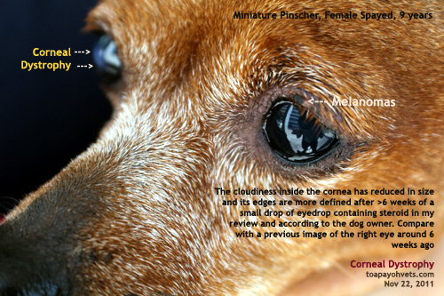
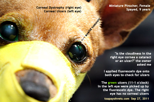
Use digital images from a good camera lens able to zoom to review eye cases later. In this case, the digital images picked up three very small melanomas that I missed during examination of the moving Miniature Pinscher |
||
The "melanomas"
were small but could be seen clearly in the image.
This shows that visual aids are good for reviews and
help to diagnose. The right eye is likely to suffer
from a condition called corneal endothelial dystrophy
which is a opacity (lipid deposits) of the inner layer
of the cornea due to aging.
I phoned the owner and told her about the eyelid
melanomas which can be removed by surgery if she
wished. Also, the corneal endothelial dystrophy may
develop into an ulcer later. A blood test to check for
hypothyroidism and hypercholesterol and lipids and a
low fat diet of less than 8% had been suggested. She
said that her dog is on a vegetarian diet and thanked
me for the call. Later, I e-mailed to her two relevant
images.
|
PART 3. Sep 28, 2011 Dear Ms ... I am Dr Sing from Toa Payoh Vets. Thank you for your email. Attached is a brief report and 2 images of your dog's eye conditions to help you understand what is happening to your dog's eyes. 1.
right eye - corneal endothelial dystrophy
associated with aging. It is likely due to the
leakage of the aqueous from inside the eye into
the endothelium and into the inside of the
cornea substance. Observe for corneal ulceration
which may occur later. Lipid deposit into the
cornea is also likely to cause axial corneal
opacity.2.
left eye - corneal ulcers (dyed green by
fluorescein) at around 11-1 o'clock due to the
dog's attempt to rub off the 3 eyelid tumours,
probably melanomas. The dog's eye ulceration is
known as superficial keratitis. It is not
serious presently. If the dog does not rub her
eyes (24-hour e-collar for around 4 weeks), the
ulcer will heal. My advice is to get the suspected 3 "melanomas" excised. If you wish to know whether they are cancerous or not, a histopathology done on the tumours by the laboratory will be required. Anaesthesia in any dog including old ones carry a risk of heart failure. Blood test to screen for her health before anaesthesia is advised. If you can't view the eyelid tumours, let me know and I will crop the picture focusing at the left eye. Best wishes. |